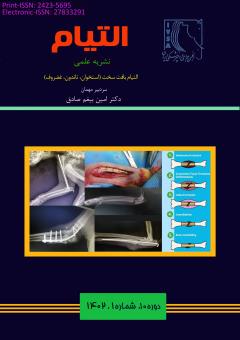
تثبیت کننده های اسکلتی خارجی در دام کوچک
محورهای موضوعی : علوم جراحی دامپزشکی شامل جراحی های بافت های سخت و نرمحمیدرضا مسلمی 1 , نوید احسانی پور 2 , فائزه عمارلو 3
1 - استادیار، گروه علوم درمانگاهی، دانشکده دامپزشکی، دانشگاه سمنان، سمنان، ایران
2 - دانشجو، گروه علوم درمانگاهی، دانشکده دامپزشکی، دانشگاه سمنان، سمنان، ایران
3 - دانشجو، گروه علوم درمانگاهی، دانشکده دامپزشکی، دانشگاه سمنان، سمنان، ایران
کلید واژه: تثبیت کننده اسکلتی خارجی, خطی, حلقوی, هیبریدی, دام کوچک,
چکیده مقاله :
تثبیت کننده های اسکلتی خارجی به عنوان یک از روش های جراحی ارتوپدی هستند که برای درمان شکستگی های باز یا بسته در استخوان های بلند، جوش دادن مفاصل، افزایش طول استخوان و ناهنجاری های مادرزادی استفاده می شوند. تثبیت کننده اسکلتی خارجی وسیله اي است که در خارج عضو قرار داده می شود و با استفاده از پین هایی که در قطعات شکسته استخوان قرار می دهند شکستگی را جا انداخته و بی حرکت می نمایند و سپس وضعیت پین ها را با اتصال به یک چهارجوب و با استفاده از پیچ و مهره هایی ثابت می کنند و بدین ترتیب قطعات شکسته در امتداد صحیح خود ثابت می شوند. اگر چه فیکساتور ها از لحاظ ظاهری و بیومکانیکی در طول زمان بسیار تغییر کرده اند اما دارای اصول و کارکرد یکسانی هستند. به گونه ای که این فیکساتور ها از یک پین یا سیم های نازک از جنس استیل ضد زنگ تشکیل شده اند که داخل پوست فرو رفته و به استخوان می رسد. این فیکساتور ها بر اساس ساختار هندسی پیکره و قالب انواع مختلفی دارند که شامل فیکساتور های خطی، دایره ای و هیبریدی می -باشد. ساده ترین و پرکاربرد ترین نوع فیکساتور های اسکلتی خارجی، فیکساتور های خطی هستند. استفاده از این روش ها نسبت به روش های دیگر دارای مزایایی از جمله پایدارکردن شکستگی استخوان با فاصله از محل صدمه، عدم وجود گچ، حرکت راحت بیمار، دخالت مفصل به میزان کم می باشد. شل شدن زودرس پین ها، شایع ترین عارضه است که سبب درد، التهاب و ترشح از مجرای پین می شود. این فیکساتورها یک مدل درمانی چندوجهی و موثر است، اما نیاز به مراقبت دقیق در طول دوره درمان دارد. قبل از تصمیم گیری برای استفاده از فیکساتورهای خارجی، باید امکان رعایت دستورالعمل های مراقبت پس از عمل توسط صاحبان بیمار و حیوانات خانگی در نظر گرفته شود. در این مقاله به بررسی انواع فیکساتور های اسکلتی خارجی و مدیریت آنها بعد از جراحی و عوارض آن پرداخته می شود.
An external skeletal fixator is an orthopedic method for treating open or closed fractures of long tubular bones, joint stiffness, bone lengthening, and congenital malformations. An external skeletal fixator is a device that is installed outside the organ and inserts pins into the fracture to fix it and adjust the position of the pin. They are connected to the frame and secured with bolts and nuts. Fixtures have changed significantly in appearance and biomechanics over time, but the principle and function remain the same. These fixtures consist of pins or thin stainless steel wires that penetrate the skin and reach the bone. This way the broken part is fixed in the right direction. Depending on the body geometry and shape, these external skeletal fixators are available in different types such as linear, circular, and hybrid fixators. The simplest and most common type of external skeletal fixator is the linear fixator. The use of an external fixator has several advantages over other fixation methods such as stabilization of the fracture at some distance from the injury site, no need for a cast, ease of patient movement, and minimal involvement of the joint. Premature loosening of the pin is the most common complication causing pain, inflammation, and discharge from the pin tract. Although these fixators are versatile and effective treatment models, they require careful maintenance during treatment. Before deciding to use an external fixator, the patient's and pet's owner's ability to comply with postoperative care instructions should be considered. This article reviews the types of external fixators, postoperative care, and their complications.
1. Cinthuja P, Wijesinghe PCI, Silva P. Use of external fixators in developing countries: a short socioeconomic analysis. Cost Effectiveness and Resource Allocation. 2022;20(1):1-9.
2. Mathieu L, Ouattara N, Poichotte A, Saint-Macari E, Barbier O, Rongiéras F, et al. Temporary and definitive external fixation of war injuries: use of a French dedicated fixator. International orthopaedics. 2014;38:1569-76.
3. Thomas SR, Giele H, Simpson AH. Advantages and disadvantages of pinless external fixation. Injury. 2000;31(10):805-9.
4. Bakici M, Karsl B, Cebeci MT. External skeletal fixation. International Journal of Veterinary and Animal Research. 2019;2(3):69-73.
5. Fossum TW, Cho J, Dewey CW, Hayashi K, Huntingford JL, MacPhail CM, et al. Small animal surgery. Philadelphia, PA : Elsevier, Inc. 2019.
6. Sylvestre AM. Fracture Management for the Small Animal Practitioner. John Wiley & Sons, Inc. 2019.
7. Mathew DD, Ranganath L, Nagaraja B. Comparison of Type Ia single and double connecting bar external skeletal fixation for femoral fracture repair in dogs. Indian Journal of Veterinary Surgery. 2010;31(2):133-4.
8. Pardeshi G, Ranganath L. Comparison of type Ia and type Ib external skeletal fixation for tibial fracture repair in dogs. Indian Journal of Veterinary Surgery. 2008;29(2):93-5.
9. Corr SA. Practical guide to linear external skeletal fixation in small animals. In Practice. 2005;27: 76-85.
10. Egger EL. External Skeletal Fixation. In: Bojrab MJ (Ed), Current Techniques in Small Animal Surgery. Williams & Wilkins, Maryland. 1998.
11. Fernando P, Abeygunawardane A, Wijesinghe P, Dharmaratne P, Silva P. An engineering review of external fixators. Medical Engineering and Physics. 2021;98:91-103.
12. Kirkby KA, Lewis DD, Lafuente MP, Radasch RM, Fitzpatrick N, Farese JP, et al. Management of humeral and femoral fractures in dogs and cats with linear-circular hybrid external skeletal fixators. Journal of the American Animal Hospital Association. 2008;44(4):180-97.
13. Hudson CC, Lewis DD, Cross AR, Dunbar NJ, Horodyski M, Banks SA, et al. A Biomechanical Comparison of Three Hybrid Linear-Circular External Fixator Constructs. Veterinary Surgery. 2012;41(8):954-65.
14. Jiménez-Heras M, Rovesti GL, Nocco G, Barilli M. Evaluation of sixty-eight cases of fracture stabilisation by external hybrid fixation and a proposal for hybrid construct classification. BMC Veterinary Research. 2014;10(1):1-10.
15. Beever LJ, Giles K, Meeson RL. Postoperative Complications Associated with External Skeletal Fixators in Dogs. Veterinary and Comparative Orthopaedics and Traumatology. 2018;31(2):137-43.


