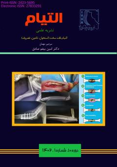
روش¬های تثبیت داخلی شکستگی¬های استخوان در دام¬های کوچک
محورهای موضوعی : Veterinary Soft and Hard Tissue Surgery
آرین پورامین
1
![]() ,
سیف الله دهقانی ناژوانی
2
,
سیف الله دهقانی ناژوانی
2
1 - دانشجو، گروه علوم درمانگاهی، دانشکده دامپزشکی، دانشگاه شیراز، شیراز، ایران
2 - استاد، گروه علوم درمانگاهی، دانشکده دامپزشکی، دانشگاه شیراز، شیراز، ایران
کلید واژه: شکستگی, استخوان, تثبیت داخلی, پین, پیچ و پلاک,
چکیده مقاله :
شکستگی استخوان ها در سگ ها و گربه ها معمولا جزء مواردی است که در کلینیک می بینیم و با آن سر و کار داریم. در حالی که معمولا شکستگیها پس از یک حادثه آسیبزا مانند برخورد با ماشین یا افتادن از ارتفاع رخ میدهند، برخی از شکستگیها به دنبال ضعیف شدن پاتولوژیک استخوان رخ میدهند که با برخی شرایط نئوپلاستیک مانند استئوسارکوم دیده میشود. اهداف اصلی تثبیت شکستگی ها؛ بی حرکت سازی قطعات شکستگی، ترمیم سریع استخوان آسیب دیده، بازگشت تحرک اولیه و عملکرد کامل اندام آسیب دیده و ثبات برای تحمل وزن توسط اندام حرکتی آسیب دیده می باشد. تمام روشهای تثبیت داخلی و خارجی که امکان حرکت بین قطعه ای قابلتوجهی را تحت تحمل وزن فراهم میکنند، به عنوان تثبیت انعطافپذیر در نظر گرفته میشوند. نکته قابل توجه در تثبیت شکستگی ها این است که امکان ترمیم وجود داشته باشد یعنی خون رسانی به محل حفظ شود و تثبیت آنقدر سفت نشود که منجر به تاخیر در جوش خوردگی استخوان شود. جا اندازی باز و تثبیت داخلی برای بازگرداندن آناتومی استخوان و تحرک اولیه و برای غلبه بر محدودیتهایی که هنگام درمان شکستگیها با کشش اسکلتی یا بیحرکتی گچ گرفته میشوند، استفاده شده است. هدف اصلی تثبیت داخلی دستیابی سریع و در صورت امکان عملکرد کامل اندام آسیب دیده با توانبخشی سریع بیمار است. انتخاب روش تثبیت داخلی بر اساس طبقه بندی شکستگی، استخوان آسیب دیده، آسیب های همزمان، شکستگی باز در مقابل بسته و البته نیروهایی است که با روش تثبیت خنثی می شوند. ایمپلنت های تثبیت داخلی از جنس استیل ضد زنگ هستند و شامل؛ پین داخل مدولای استخوان، سیم ارتوپدی، پیچ و پلاک می باشند.
Bone fractures in dogs and cats are usually see and we concern with those in the clinic. Usually, fractures occur after a traumatic accident such as being hit by a car or falling from a height, some fractures occur following pathological bone weakening, which is seen with some neoplastic conditions such as osteosarcoma. The main goals of fracture stabilization; Immobilization of broken parts, quick repair of the damaged bone, return of primary mobility, full function and stability to weight bearing of the bruise limb. All internal and external fixation methods that allow significant intersegmental motion under weight bearing are considered flexible fixation. The best important point in the stabilization of fractures is that a possibility of repair, that is, the blood supply to the place is maintained and the fixation is not so tight that it leads to a delay in bone fusion. Open fixation and internal fixation have been used to restore bone anatomy and original mobility and to overcome the limitations encountered when treating fractures with skeletal traction or cast immobilization. The main goal of internal fixation is to achieve rapid and, if possible, full function of the affected limb with rapid rehabilitation of the patient. The selection of the internal fixation method is based on the classification of the fracture, break bone, synchronize injuries, open fracture, and of course the forces that are neutralized by the fixation method. Internal stabilization implants are made of stainless steel and include; There are intramedullary pins, orthopedic wire, plates and screws.
1. Ruedi TP, Murphy WM, eds. AO principles of fracture management. Stuttgart, Germany: Thieme, 2000.
2. Benjamin BB, Lund PJ. Orthopedic devices. In: Hunter TB, Bragg DG, eds. Radiographic guide to medical devices and foreign bodies. St Louis, Mo: Mosby–Year Book, 1994; 348–385.
3. Wiss DA, ed. Fractures. In: Thompson RC, ed. Master techniques in orthopedic surgery. Philadelphia, Pa: Lippincott-Raven, 1998.
4. Berquist TH, ed. Imaging atlas of orthopedic appliances and prostheses. New York, NY: Raven, 1995.
5. Freiberg AA, ed. The radiology of orthopedic implants: an atlas of techniques and assessment. St Louis, Mo: Mosby, 2001.
6. Hunter TB, Taljanovic M. Overview of medical devices. Curr Probl Diagn Radiol 2001; 30:94–139.
7. Taljanovic, M. S., Jones, M. D., Ruth, J. T., Benjamin, J. B., Sheppard, J. E., & Hunter, T. B. (2003). Fracture fixation. Radiographics, 23(6), 1569-1590.
8. Johnston SA, von Pfeil DJF, Dejardin LM, et al. Internal fracture fixation. In Tobias KM, Johnston SA (eds): Veterinary Surgery, Small Animal, 1st ed. Philadelphia: Elsevier, 2012.
9. Harasen G. Orthopedic hardware and equipment for the beginner: Part 1. Pins and wires. Can Vet J 2011; 52(9):1025-1026.
10. Martinez SA, DeCamp CE. External skeletal fixation. In Tobias KM, Johnston SA. Veterinary Surgery, Small Animal, 1st ed. Philadelphia: Elsevier, 2012.


